

NIR-II IN VIVO Imaging
Small animal Imaging in the Second Near-Infrared (NIR-II) Window
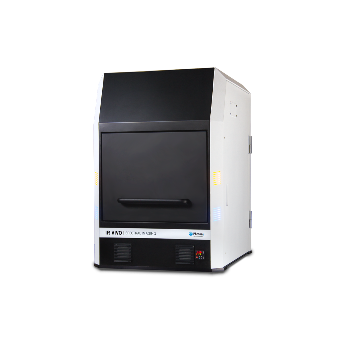
The IR VIVO family stands as a transformative breakthrough in the realm of small animal optical imaging.
Empower your research endeavors by gaining the ability to visualize biomarkers in their dynamic in vivo context, all in real-time. With a keen focus on optimizing contrast resolution and sensitivity, IR VIVO utilizes state-of-the-art NIR-II technology, anchored by Photon etc. ultra-low-noise InGaAs camera (Alizé 1.7 or ZephIR 1.7). Complemented by novel homogeneous illumination and robust analytical software, this system delivers an unparalleled fusion of rapid, high-resolution, and profound imaging capabilities, cementing its role as a monumental advancement in the bioluminescence imaging field of small animal optical imaging system for driving significant strides in biodistribution studies and cancer research.
Real-Time Biodistribution of ICG in a Mouse Using NIR-II Fluorescence Imaging
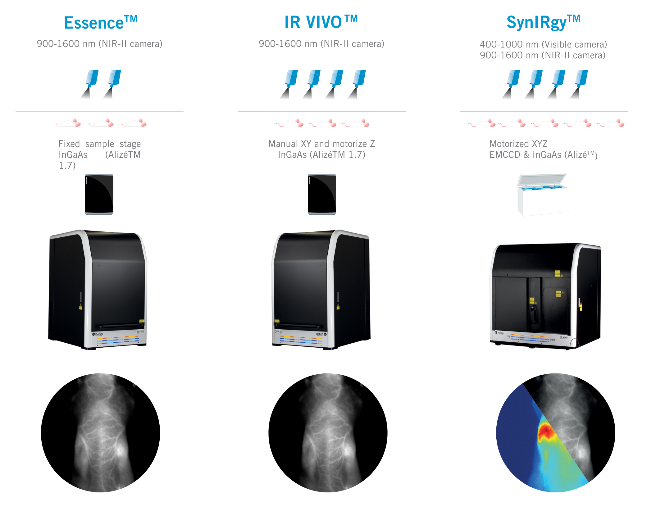
The IR VIVO family has three flavors:
- IR Vivo: Experience the pinnacle of small animal fluorescence imaging with the state-of-the-art IR Vivo model, the standard system for the NIR-II field. Elevate your research with unparalleled sensitivity, resolution, and imaging capabilities.
- Essence: Simplify your journey into in vivo imaging with the Essence model. Streamline your research endeavors, achieve swift and clear insights without compromising on quality.
- SynIRgy: Enter a new era of fluorescence imaging synergy with the SynIRgy model. Harness the combined power of near-infrared ranges and bioluminescence for a comprehensive biomarker visualization and achieve a perfect balance of resolution and sensitivity for groundbreaking biodistribution studies and cancer research advancements.
Product Specifications
| IR VIVO | Essence | SynIRgy | |
|---|---|---|---|
| Emission spectral range | 900-1600 nm | 900-1600 nm | 400-1000 nm (Visible camera) 900-1600 nm (NIR-II camera) |
| Filtering | Filter wheel with up to 6 emission channels | Filter wheel with up to 6 emission channels | Filter wheel with up to 6 emission channels |
| Illumination source | Laser at 670, 760, 808 and 890 nm and adjustable power density | Laser at 808 and 890 nm and adjustable power density | Laser at 465, 520, 670, 760 et 808 nm and adjustable power density |
| Lens | 50mm f/1.4 lens | 50mm f/1.4 lens | VIS: 35mm f/0.95 NIR-II: 50mm f/1.4 |
| Field of view | 80 x 64 mm to 50 x 40mm Variable FOV for 1 mouse or individual organ view | Fixed FOV for 1 mouse imaging: 80 x 64 mm | 80 x 64 mm to 50 x 40mm Variable FOV for 1 mouse or individual organ view |
| Stage | Manual XY and motorize Z | Fixed sample stage | Motorized XYZ |
| Dimensions ( L x W x H) | 77 x 60 x 98 cm | 77 x 60 x 98 cm | 92 x 76 x 110 cm |
| Camera type | InGaAs (Alizé™ 1.7) | InGaAs (Alizé™ 1.7) | EMCCD & InGaAs (Alizé™ 1.7) |
Spec Sheet
Applications
Publications
Journal of Biomedical Optics
- Biochemistry & Nanosensors
Minimizing near-infrared autofluorescence in preclinical imaging with diet and wavelength selection
Journal of Nanobiotechnology
- Preclinical
In vivo fluorescence imaging: success in preclinical imaging paves the way for clinical applications
Journal of BioPhotonics
- Preclinical
The near-infrared autofluorescence fingerprint of the brain
Nature Communications
- Preclinical
Ultrafast photochemistry produces superbright short-wave infrared dots for low-dose in vivo imaging
Expert Opinion on Drug Discovery
- Pharmacology
In vivo near-infrared fluorescent optical imaging for CNS drug discovery
PloS one
- Carbon-Based Materials
- Biochemistry & Nanosensors
Long-Term in Vivo Biocompatibility of Single-Walled Carbon Nanotubes
Nano Letters
- Carbon-Based Materials
- Biochemistry & Nanosensors
- Oncology
An in Vivo Nanosensor Measures Compartmental Doxorubicin Exposure
Science Translational Medicine
- Carbon-Based Materials
- Biochemistry & Nanosensors
An Optical Nanoreporter of Endolysosomal Lipid Accumulation Reveals Enduring Effects of Diet on Hepatic Macrophages in Vivo
Science Advances
- Biochemistry & Nanosensors
- Oncology
Noninvasive Ovarian Cancer Biomarker Detection via an Optical Nanosensor Implant
Videos
IR VIVO Guided Tour
IR VIVO is an infrared multispectral imager for small animal studies. This system benefits from reduced light scattering, absorption and auto-fluorescence by using a detection system in the near and short-wave infrared. Visualize your biomarkers in vivo and in real-time with optimized contrast resolution and sensitivity. IR VIVO takes advantage of the most recent developments in SWIR imaging with an ultra-low-noise InGaAs camera (Alizé 1.7 or ZephIR 1.7), novel homogeneous illumination and powerful analytical software to provide an unprecedented combination of fast, high resolution and deep imaging.
In Vivo NIR-II Imaging: Contact-Free Monitoring Heartbeat & Respiratory Rate
High-speed imaging made available by the fast frame rates of InGaAs cameras allows for contact-free cardiography and respiratory rate measurements in both anesthetized and awake animals. Such contact-free measurements of awake mice obtained without the use of any restraining device could drastically change studies procedures in labs.
Real-Time Biodistribution and Pharmacokinetics of ICG in a Mouse Using NIR-II Fluorescence
Video of IR VIVO imaging experiment showing the biodistribution and pharmacokinetics of ICG in real time. IR VIVO is able to visualize NIR probes at high resolutions. From one single injection, it was possible to obtain measurements of heartbeats, respiration and intestinal contraction rates.
In Vivo NIR-II Imaging of Liver Metabolism, Hepatobiliary Elimination & Gastro-Intestinal Motility
With a simple tail-vein injection of ICG, we can study liver metabolism, measure hepatobiliary elimination and monitor gastrointestinal motility simultaneously in vivo and contact-free. Such a straightforward method to image the intestine could help research on metabolic diseases.
White Papers
In Vivo NIR-II Imaging: A Disruptive Force for Preclinical Research
Authors Stephen Marchant, Jacob Yvon-Leroux and Émilie Beaulieu Ouellet